Call 830-379-3937

Glaucoma care · San Antonio

Glaucoma treatment and MIGS surgery in San Antonio

Glaucoma damages the optic nerve, usually because fluid inside the eye doesn't drain well and pressure builds. Every glaucoma treatment works by lowering that pressure, either by making less fluid or by helping it drain. As a fellowship-trained glaucoma specialist, Dr. Kavanagh has performed every type of MIGS procedure and traditional glaucoma surgery, so your treatment is matched to your glaucoma rather than to the tools a surgeon happens to use.

CORNEAANTERIOR CHAMBERIRIS LENSSCLERACILIARY BODY CONJUNCTIVA THE DRAINAGE ANGLE - - > FLUID OUTFLOW ROUTE
Fluid leaves the eye through the drainage angle. Each procedure below improves a different part of this system.

Jump to a procedure

Compare glaucoma procedures

ProcedureTypeWhere it worksSettingWith cataract surgery?
SLT laserLaserTrabecular meshwork (natural drain)In office, about 5–10 minutesNot needed
DurystaDrug implantInside the eye's front chamberOffice procedureNot needed
iDose TRDrug implantAnchored in the drainage angleSurgery centerCan be done alone or with cataract surgery
iStent infiniteMIGS stentTrabecular meshwork into Schlemm's canalSurgery centerCan be done alone or with cataract surgery
Hydrus MicrostentMIGS stentSchlemm's canal (scaffolds about 90 degrees)Surgery centerPerformed with cataract surgery
AlloFlo UveoMIGS, uveoscleralSupraciliary space (uveoscleral pathway)Surgery centerPerformed as a standalone procedure
XEN Gel StentMIGS, subconjunctivalFrom the front chamber to under the conjunctivaSurgery centerCan be done alone or with cataract surgery
TrabeculectomyTraditional glaucoma surgeryNew drain under a scleral flap into a blebSurgery center or hospitalUsually separate
Ahmed valve & seton surgeryTube shuntTube in the eye drains to a plate on the scleraSurgery center or hospitalUsually separate

For educational purposes. The right procedure depends on your type and stage of glaucoma.

CORNEAANTERIOR CHAMBERIRIS LENSSCLERACILIARY BODY CONJUNCTIVA SLT LASER - - > FLUID OUTFLOW ROUTE

Where it works: Trabecular meshwork (natural drain)
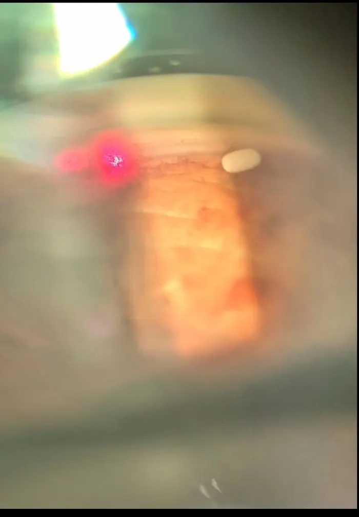
View through a gonioscopy lens during SLT laser, with the red aiming beam on the drainage angle
LaserIn office, about 5–10 minutes

Selective laser trabeculoplasty (SLT)

SLT uses short pulses of low-energy laser light on the trabecular meshwork, the eye's natural drain. The laser stimulates the body's own repair process so fluid drains more easily.

The procedure

  • Numbing drops are placed and a special contact lens rests on the eye
  • The laser is applied around the drainage angle in a few minutes
  • You go home the same day and resume normal activities

Often a good fit for

Newly diagnosed open-angle glaucoma, patients who want to reduce or avoid daily drops, and those who have trouble with drops or their side effects. Large studies support SLT as a first-line treatment.

The effect can fade over years, and SLT can be repeated.
CORNEAANTERIOR CHAMBERIRIS LENSSCLERACILIARY BODY CONJUNCTIVA DURYSTA - - > FLUID OUTFLOW ROUTE

Where it works: Inside the eye's front chamber

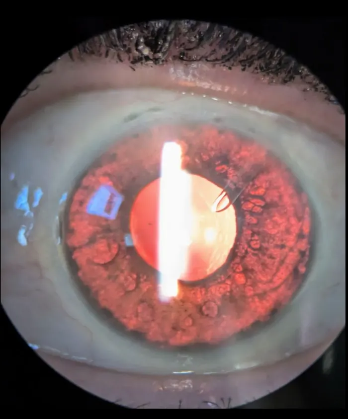
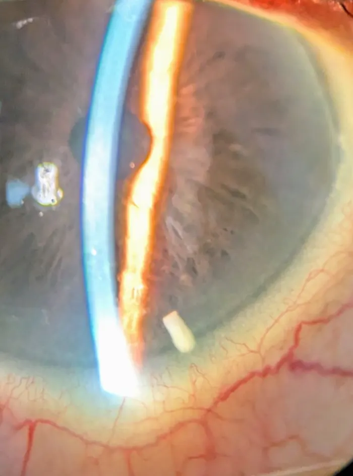
A Durysta implant resting in the drainage angle of the eye
Drug implantOffice procedure

Durysta (bimatoprost implant)

Durysta is a tiny, dissolvable implant that slowly releases bimatoprost, a common glaucoma medication, from inside the eye. It lowers pressure without daily drops for months in many patients.

The procedure

  • The eye is numbed and cleaned
  • The implant is placed in the front chamber with a fine applicator
  • The implant gradually dissolves as it releases medication

Often a good fit for

Patients with open-angle glaucoma or ocular hypertension who struggle with daily drops, forget doses or have drop side effects.

Not suitable for every eye, including some with corneal problems. Dr. Kavanagh checks your cornea first.

CORNEAANTERIOR CHAMBERIRIS LENSSCLERACILIARY BODY CONJUNCTIVA IDOSE TR - - > FLUID OUTFLOW ROUTE

Where it works: Anchored in the drainage angle

Gonioscopic view of an iDose TR implant anchored in the drainage angle
Drug implantSurgery center

iDose TR (travoprost intracameral implant)

iDose TR is a tiny titanium implant anchored in the eye's drainage tissue. It continuously releases travoprost, a proven glaucoma medication, directly inside the eye for an extended period, reducing or eliminating the need for daily drops.

The procedure

  • Performed through a tiny incision at the edge of the cornea
  • The implant is anchored in the wall of the drainage angle
  • It can be placed during cataract surgery or as its own procedure

Often a good fit for

Patients with open-angle glaucoma or ocular hypertension who want a long-lasting alternative to daily drops.

It treats with medication rather than creating a new drain, so it pairs well with MIGS stents.

CORNEAANTERIOR CHAMBERIRIS LENSSCLERACILIARY BODY CONJUNCTIVA ISTENT INFINITE - - > FLUID OUTFLOW ROUTE

Where it works: Trabecular meshwork into Schlemm's canal

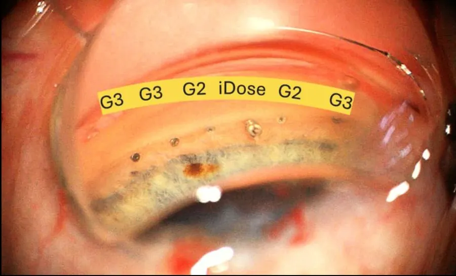
Gonioscopic view showing iStent infinite stents alongside an iDose TR implant
MIGS stentSurgery center

iStent infinite trabecular micro-bypass

iStent infinite places three heparin-coated titanium micro-stents through the trabecular meshwork into Schlemm's canal. Each stent opens a bypass through the blocked drain, and spreading three stents around the angle reaches more of the eye's collector channels.

The procedure

  • Performed through a tiny corneal incision using a microscope and gonioscopy lens
  • A preloaded injector places three stents in one pass
  • Recovery is quick, usually with drops for a few weeks

Often a good fit for

Open-angle glaucoma not controlled by medication or prior surgery. It is FDA-approved as a standalone procedure, so it does not require cataract surgery.

The stents are among the smallest implants in medicine; you will not feel them.

CORNEAANTERIOR CHAMBERIRIS LENSSCLERACILIARY BODY CONJUNCTIVA HYDRUS MICROSTENT - - > FLUID OUTFLOW ROUTE

Where it works: Schlemm's canal (scaffolds about 90 degrees)

MIGS stentSurgery center

Hydrus Microstent

The Hydrus is an 8 mm flexible nitinol scaffold that sits inside Schlemm's canal, holding the canal open across roughly a quarter of the eye's circumference while bypassing the trabecular meshwork. Dr. Kavanagh was the first surgeon in South Texas to implant the Hydrus.

The procedure

  • Placed through the same small incision used for cataract surgery
  • The scaffold is guided into Schlemm's canal under direct view
  • Healing is typically quick, similar to cataract surgery alone

Often a good fit for

Mild to moderate open-angle glaucoma in patients having cataract surgery. Long-term studies show lower pressure and fewer medications compared with cataract surgery alone.

Because it is placed with cataract surgery, it adds little recovery time.

CORNEAANTERIOR CHAMBERIRIS LENSSCLERACILIARY BODY CONJUNCTIVA ALLOFLO UVEO - - > FLUID OUTFLOW ROUTE

Where it works: Supraciliary space (uveoscleral pathway)

MIGS, uveoscleralSurgery center

AlloFlo Uveo (supraciliary bio-tissue spacer)

Most MIGS procedures work on the trabecular drain. AlloFlo Uveo works on the eye's other natural outflow route, the uveoscleral pathway. A small, controlled opening is made between the ciliary body and the sclera, and tiny spacers made of processed donor scleral tissue are placed to keep that space open so fluid can drain.

The procedure

  • Performed as its own procedure, separate from cataract surgery
  • Done through a small incision from inside the eye
  • A gentle separation is created between the ciliary body and the sclera (a cyclodialysis)
  • Donor-tissue spacers are inserted to keep the new pathway open

Often a good fit for

Open-angle glaucoma, including patients whose earlier trabecular procedures have lost effect.

AlloFlo uses natural, collagen-rich tissue rather than a synthetic implant.

CORNEAANTERIOR CHAMBERIRIS LENSSCLERACILIARY BODY CONJUNCTIVA XEN GEL STENT - - > FLUID OUTFLOW ROUTE

Where it works: From the front chamber to under the conjunctiva

MIGS, subconjunctivalSurgery center

XEN Gel Stent

The XEN Gel Stent is a soft, 6 mm tube made of gelatin that creates a new drainage channel from inside the eye to the space under the conjunctiva, the clear skin over the white of the eye. Fluid collects there in a small reservoir called a bleb and is absorbed. It lowers pressure more than trabecular MIGS, with less tissue disruption than a trabeculectomy.

The procedure

  • The stent is placed through a tiny incision, usually from inside the eye
  • A medication (mitomycin C) is used to reduce scarring
  • Follow-up visits are more frequent early on, and some eyes need a minor in-office adjustment of the bleb

Often a good fit for

Glaucoma that remains uncontrolled despite medications or earlier surgery, when a lower target pressure is needed.

Because it forms a bleb, you will have more follow-up visits in the first months.

CORNEAANTERIOR CHAMBERIRIS LENSSCLERACILIARY BODY CONJUNCTIVA TRABECULECTOMY - - > FLUID OUTFLOW ROUTE

Where it works: New drain under a scleral flap into a bleb

Traditional glaucoma surgerySurgery center or hospital

Trabeculectomy

Trabeculectomy has been the gold standard for lowering eye pressure for decades. A small, protected opening is created in the wall of the eye under a trap-door flap of sclera. Fluid drains through it into a bleb under the conjunctiva, often achieving the lowest pressures of any glaucoma surgery.

The procedure

  • Performed in the operating room with anesthesia so you are comfortable
  • A partial-thickness flap is made in the sclera and a small opening is created beneath it
  • Sutures control the flow and can be adjusted after surgery; frequent follow-up is needed early on

Often a good fit for

Advanced or rapidly progressing glaucoma, and eyes needing very low pressure.

Lifelong awareness matters: call right away for a red, painful eye or discharge after a trabeculectomy.

CORNEAANTERIOR CHAMBERIRIS LENSSCLERACILIARY BODY CONJUNCTIVA AHMED VALVE & SETON SURGERY - - > FLUID OUTFLOW ROUTE

Where it works: Tube in the eye drains to a plate on the sclera

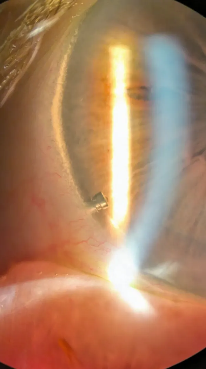
An eye with an Ahmed glaucoma valve tube visible inside the front chamber
Tube shuntSurgery center or hospital

Ahmed glaucoma valve and tube shunts (seton surgery)

A tube shunt, also called a seton, places a thin silicone tube into the eye that carries fluid to a small plate secured on the outer wall of the eye, behind the eyelid. The Ahmed valve includes a pressure-sensitive valve that helps prevent pressure from dropping too low right after surgery. Dr. Kavanagh was the first ophthalmologist to perform seton surgery in Seguin.

The procedure

  • Performed in the operating room
  • The plate is secured to the sclera under the conjunctiva and the tube is placed into the eye
  • The tube is covered with a patch graft for protection

Often a good fit for

Advanced, complex or hard-to-control glaucoma, including neovascular and uveitic glaucoma and eyes where earlier surgery has failed.

Many patients still use some drops afterward, usually fewer than before.

Questions

Glaucoma treatment questions

Which glaucoma procedure is right for me?

It depends on the type and stage of your glaucoma, your target pressure, whether you need cataract surgery and how well you tolerate drops. Dr. Kavanagh reviews your optic nerve scans, visual fields and pressure history to recommend a plan.

Will I still need eye drops?

Many patients use fewer drops after a procedure, and some stop entirely. The goal is the right pressure for your optic nerve with the fewest burdens.

Can glaucoma damage be reversed?

No. Vision already lost to glaucoma cannot be restored, which is why early diagnosis and steady pressure control matter so much.

What is MIGS?

Minimally invasive glaucoma surgery: procedures performed through tiny incisions, often with cataract surgery, that improve drainage with faster recovery than traditional surgery.

For educational purposes. This page explains glaucoma treatments to help you prepare for your visit. Your exam determines the right plan for your eyes.

Protect your vision. See a glaucoma specialist in San Antonio.

Call 830-379-3937 Book